Dentist with a Special Interest or Specialist – What’s the Difference?
October 21, 2026
Child Still in Pain After Root Canal? | Bristol Patient Story
November 12, 2026
CASE OF THE MONTH: What would you do?
Dental trauma: Two injured incisors, two very different treatment decisions Case presented by Dr Armineh Arzandeh, Specialist Endodontist, Stoke Lane Dentistry
A 16-year-old patient was referred with persistent pain following a traumatic injury to her upper central incisors.
What initially appeared to be a complication following emergency root canal treatment turned out to be a more complex case involving two adjacent teeth, each requiring a very different management approach.
This case highlights the importance of accurate diagnosis, appropriate intervention and knowing when monitoring may be the best course of action.

1. What would you do?
The clinical presentation
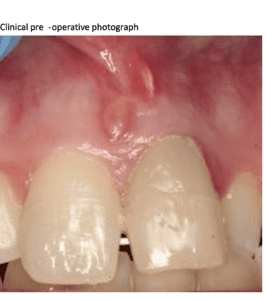
Patient, aged 16, had fallen onto her face while playing football two weeks earlier, sustaining injuries to her nose, upper lip and both maxillary central incisors (UL1 and UR1).
Following an emergency appointment with her dentist, root canal treatment was carried out on UL1 and the tooth was restored with composite.
However, UL1 remained symptomatic, and the referring dentist had noticed something unusual during treatment.
Emma was referred to Dr Armineh Arzandeh for further investigation.
The clinical challenge
You have a young patient with persistent pain following dental trauma and recent root canal treatment. The initial periapical radiograph suggests that the root filling may lie outside the root canal.
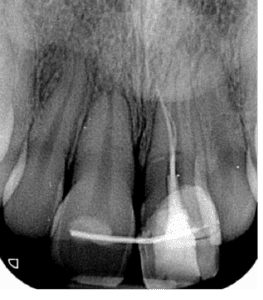
What would you do next?
-
Would you consider further imaging?
-
What complications might explain the persistent symptoms?
-
Would you investigate the adjacent incisor, even if it was asymptomatic?
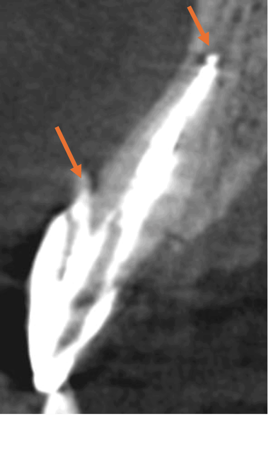
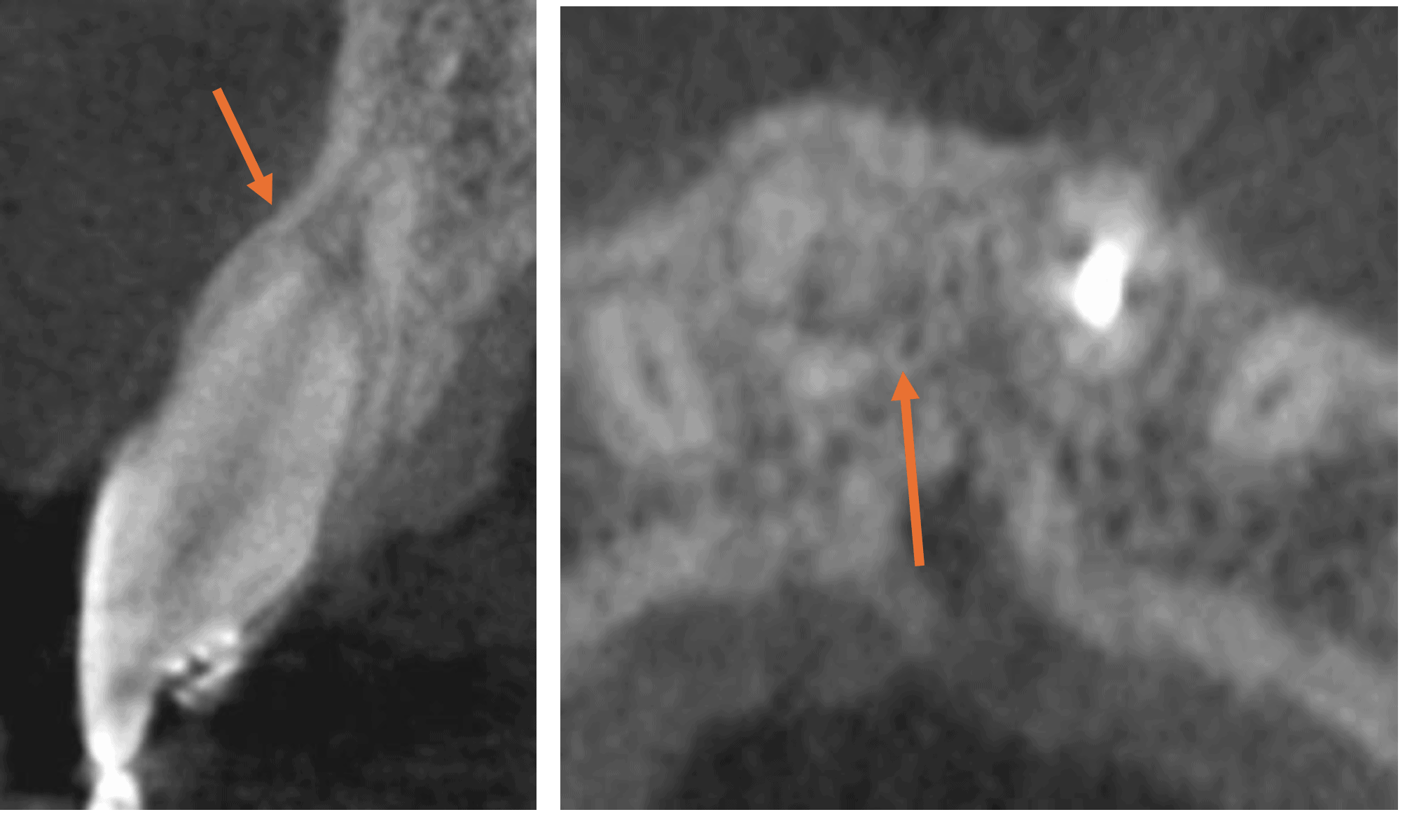
CBCT sagittal and axial views, with arrows highlighting the perforation, extruded gutta-percha and root fracture.
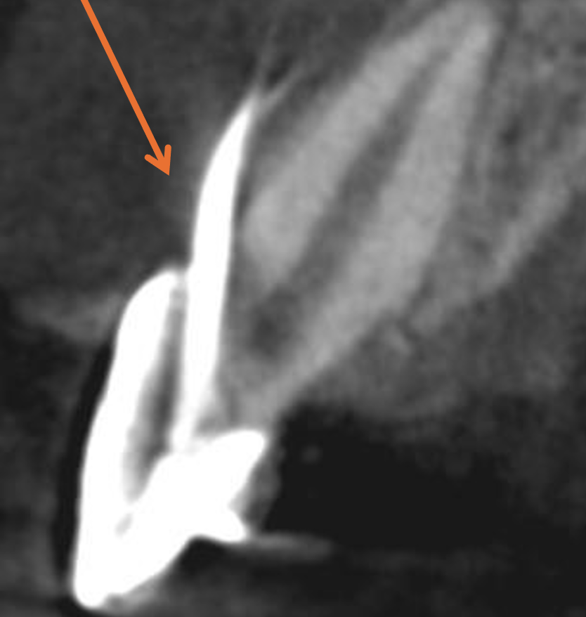
Sagittal view -1
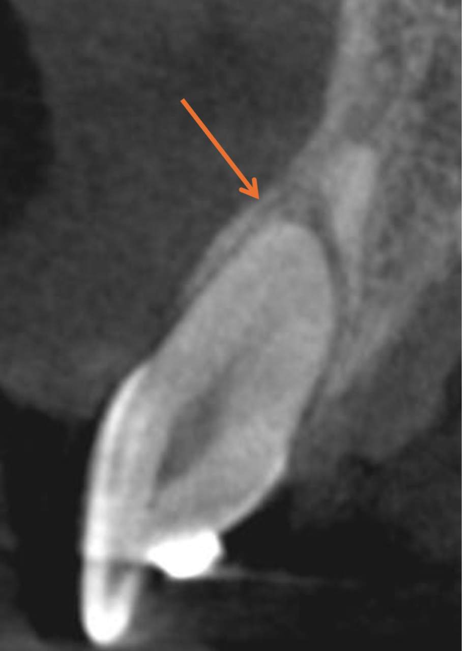
Sagittal view -2
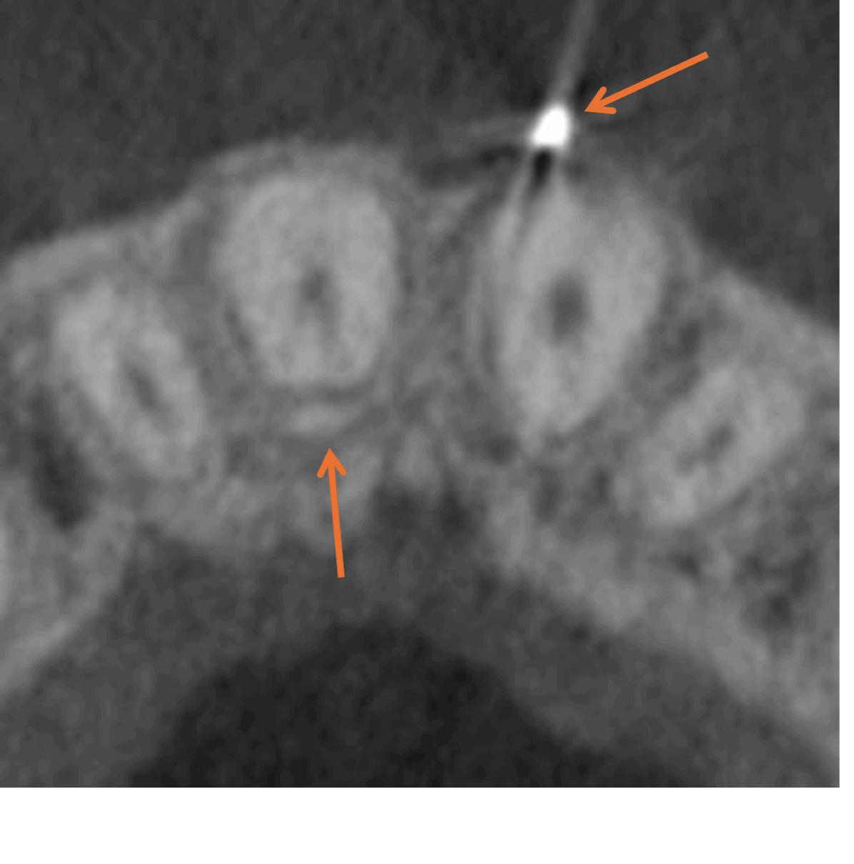
Axial view
3. What did we do – and why?
UL1 – Retreatment and perforation repair
The objective was to preserve UL1 through a non-surgical approach wherever possible.
The treatment plan involved:
-
Root canal retreatment.
-
Retrieval of the extruded gutta-percha through an orthograde approach.
-
Internal repair of the buccal perforation.
A surgical repair was discussed with Emma and her mother as an alternative should the orthograde approach prove unsuccessful.
Fortunately, all procedures were successfully completed through the orthograde approach in a single visit.
UR1 – Why no treatment was necessary
The management decision for UR1 was quite different.
Despite the horizontal root fracture, the tooth remained asymptomatic and was not mobile.
The decision was therefore made to monitor rather than intervene.
Horizontal root fractures can have a favourable prognosis, particularly when located towards the apical portion of the root.
The important distinction: One tooth required active specialist treatment, while the other could be managed conservatively through observation.
The six-month outcome
At the six-month review, both teeth showed excellent healing, demonstrating the benefits of two different treatment approaches.
UL1 – Successful endodontic retreatment
The perforation repair was satisfactory, with complete healing of the associated apical pathology. The surrounding gingival tissues had also healed well, with minimal recession.
UR1 – Healing without intervention
The adjacent incisor, which had been carefully monitored rather than treated, showed encouraging signs of natural healing. CBCT confirmed bony infill associated with the horizontal root fracture
The result? Both natural front teeth were preserved.
For a 16-year-old patient, this was a particularly positive outcome. By combining specialist intervention where necessary with a conservative approach where appropriate, both teeth could be retained without surgical treatment.
Annual reviews were planned to monitor their continued stability and long-term health.
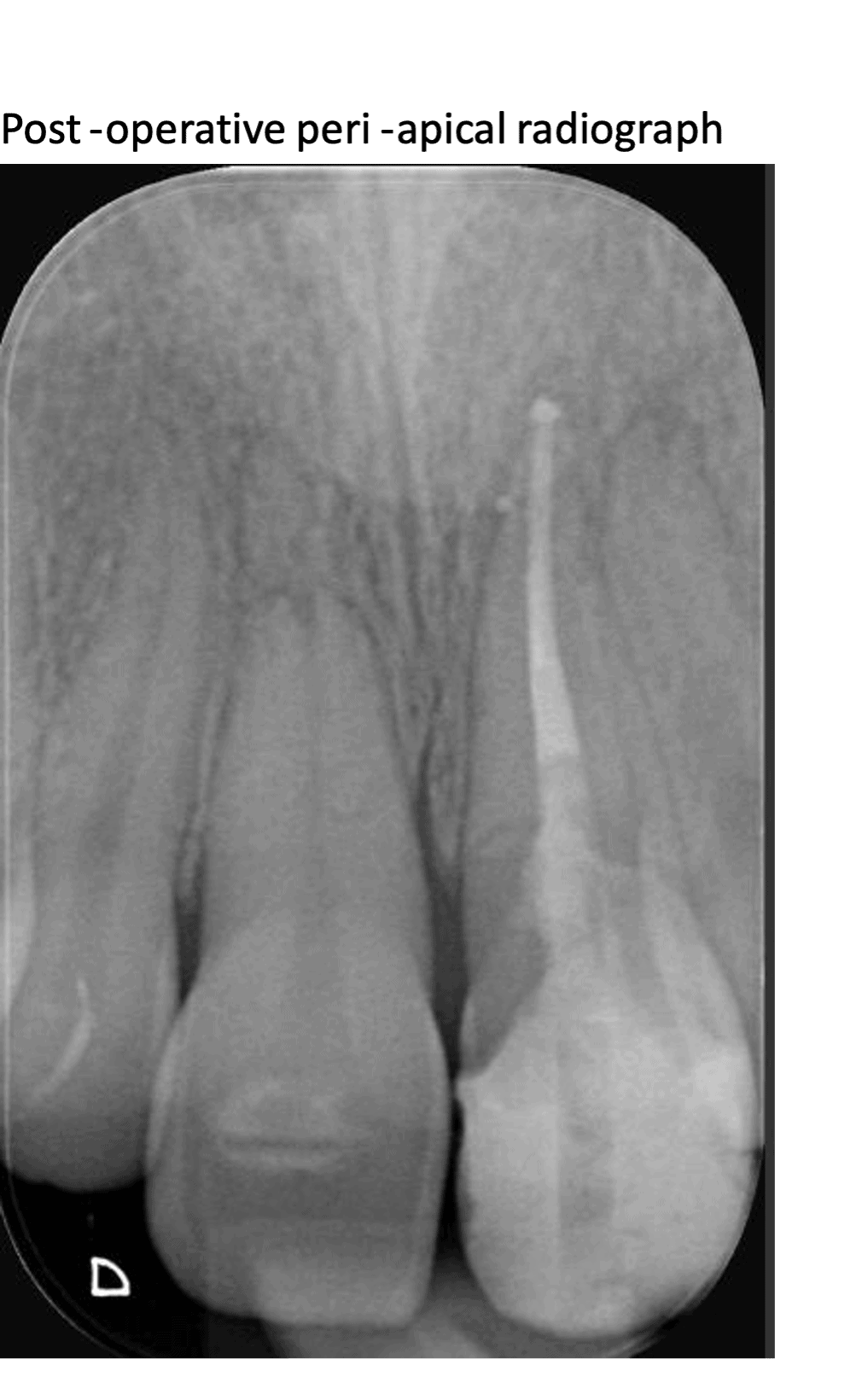
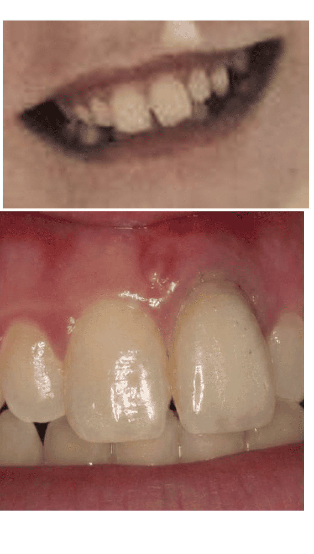
Complete healing was achieved
4. Take-home learning
Four practical lessons for general dental practice
1. Persistent symptoms warrant reassessment
When pain continues following emergency endodontic treatment, consider whether there may be an underlying complication requiring further investigation.
2. Don't overlook adjacent teeth following trauma
A patient's main complaint may relate to one tooth, but neighbouring teeth can have injuries that are initially asymptomatic.
3. Appropriate imaging can influence treatment planning
In this case, CBCT provided valuable three-dimensional information about the perforation, extruded gutta-percha and adjacent root fracture, helping guide clinical management.
4. Not every root fracture requires active treatment
Where the clinical findings are favourable, careful monitoring may be appropriate. The decision should be based on the fracture characteristics, clinical signs and ongoing review.
The key message
Successful management of dental trauma isn't simply about treating the injured tooth – it's about understanding the full extent of the injury and making the appropriate decision for each tooth.
In this case, specialist endodontic retreatment preserved one incisor, while careful monitoring allowed the adjacent fractured tooth to heal without intervention.
For a 16-year-old patient, preserving both natural central incisors was a particularly valuable outcome.